Staging Breast Cancer
If the biopsy results confirm that you have breast cancer, your doctor needs to learn the extent (stage) of the disease. The stage of your breast cancer is based on:
- the size and location of the tumor
- whether cancer has spread to nearby lymph nodes or other parts of the body
- tumor grade,
- the presence of certain biomarkers
To gather this information, the oncologist may recommend additional testing such as:
-
Bone scan: The doctor injects a small amount of a radioactive substance into a blood vessel. It travels through the bloodstream and collects in the bones. A machine called a scanner detects and measures the radiation. The scanner makes pictures of the bones. The pictures may show cancer that has spread to the bones.
-
CT scan: Doctors sometimes use CT scans to look for breast cancer that has spread to the liver or lungs. An x-ray machine linked to a computer takes a series of detailed pictures of your chest or abdomen. You may receive contrast material by injection into a blood vessel in your arm or hand. The contrast material makes abnormal areas easier to see.
-
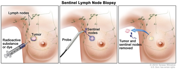
Lymph node biopsy: The stage often is not known until after surgery to remove the tumor in your breast and one or more lymph nodes under your arm. Surgeons use a method called sentinel lymph node biopsy to remove the lymph node most likely to have breast cancer cells. The surgeon injects a blue dye, a radioactive substance, or both near the breast tumor. Or the surgeon may inject a radioactive substance under the nipple. The surgeon then uses a scanner to find the sentinel lymph node containing the radioactive substance or looks for the lymph node stained with dye. The sentinel node is removed and checked for cancer cells. Cancer cells may appear first in the sentinel node before spreading to other lymph nodes and other places in the body.
The next step will be to classify the data by using a staging system. The most common system used for staging breast cancer is the TNM staging system developed by the American Joint Committee on Cancer. It describes the extent of the cancer by answering the following questions:
- Tumor (T): How large is the primary tumor in the breast? What are its biomarkers?
- Node (N): Has the tumor spread to the lymph nodes? If so, where, what size, and how many?
- Metastasis (M): Has the cancer spread to other parts of the body?
Tumor (T) Categories for Breast Cancer
T followed by a number from 0 to 4 describes the size of the primary tumor and whether it has spread to the skin or to the chest wall under the breast. Higher T numbers mean a larger tumor and/or wider spread to tissues near the breast.
TX: The primary tumor cannot be evaluated.
T0 (T zero): There is no evidence of cancer in the breast.
Tis: Refers to carcinoma in situ. The cancer is confined within the ducts of the breast tissue and has not spread into the surrounding tissue of the breast. There are 2 types of breast carcinoma in situ:
- Tis (DCIS): DCIS is a non-invasive cancer, but if not removed, it may develop into an invasive breast cancer later. DCIS means that cancer cells have been found in breast ducts and have not spread past the layer of tissue where they began.
- Tis (Paget’s disease): Paget's disease of the nipple is a rare form of early, non-invasive cancer that is only in the skin cells of the nipple. Sometimes Paget's disease is associated with invasive breast cancer. If there is an invasive breast cancer, it is classified according to the stage of the invasive tumor.
T1: The tumor in the breast is 20 millimeters (mm) or smaller in size at its widest area. This is a little less than an inch. This stage is then broken into 4 substages depending on the size of the tumor:
- T1mi is a tumor that is 1 mm or smaller.
- T1a is a tumor that is larger than 1 mm but 5 mm or smaller.
- T1b is a tumor that is larger than 5 mm but 10 mm or smaller.
- T1c is a tumor that is larger than 10 mm but 20 mm or smaller.
T2: The tumor is larger than 20 mm but not larger than 50 mm.
T3: The tumor is larger than 50 mm.
T4: The tumor falls into 1 of the following groups:
- T4a means the tumor has grown into the chest wall.
- T4b is when the tumor has grown into the skin.
- T4c is cancer that has grown into the chest wall and the skin.
- T4d is inflammatory breast cancer.
Node (N) Categories for Breast Cancer
N followed by a number from 0 to 3 indicates whether the cancer has spread to lymph nodes near the breast and, if so, how many lymph nodes are involved. Lymph nodes near where the cancer started are called regional lymph nodes. Regional lymph nodes include:
- Lymph nodes located under the arm, called the axillary lymph nodes
- Lymph nodes located above and below the collarbone
- Lymph nodes located under the breastbone, called the internal mammary lymph nodes
Lymph nodes in other parts of the body are called distant lymph nodes.
NX: The lymph nodes were not evaluated.
N0: Either of the following:
- No cancer was found in the lymph nodes.
- Only areas of cancer smaller than 0.2 mm are in the lymph nodes.
N1: The cancer has spread to 1 to 3 axillary lymph nodes and/or the internal mammary lymph nodes. If the cancer in the lymph node is larger than 0.2 mm but 2 mm or smaller, it is called "micrometastatic" (N1mi).
N2: The cancer has spread to 4 to 9 axillary lymph nodes. Or, it has spread to the internal mammary lymph nodes, but not the axillary lymph nodes.
N3: The cancer has spread to 10 or more axillary lymph nodes, or it has spread to the lymph nodes located under the clavicle, or collarbone. It may have also spread to the internal mammary lymph nodes. Cancer that has spread to the lymph nodes above the clavicle, called the supraclavicular lymph nodes, is also described as N3.
If there is cancer in the lymph nodes, knowing how many lymph nodes are involved and where they are helps doctors plan breast cancer treatment. The pathologist can find out the number of axillary lymph nodes that contain cancer after they are removed during surgery. It is not common to remove the supraclavicular or internal mammary lymph nodes during surgery. If there is cancer in these lymph nodes, treatment other than surgery, such as radiation therapy, chemotherapy, and hormonal therapy, is generally used.
Metastasis (M) Categories for Breast Cancer
Metastasis describes whether the cancer has spread to other parts of the body.
MX: Distant spread cannot be evaluated.
M0: There is no evidence of distant metastases.
M0 (i+): There is no clinical or radiographic evidence of distant metastases. However, there is microscopic evidence of tumor cells in the blood, bone marrow, or other lymph nodes that are no larger than 0.2 mm.
M1: There is evidence of metastasis to another part of the body, meaning there are breast cancer cells growing in other organs.
Breast Cancer Stage Groups
The oncologist will assign the stage of the cancer by combining the T, N, and M classifications, the tumor grade, and the results of ER/PR and HER2 testing. This information will guide your cancer care team as they develop your treatment plan and determine your prognosis.
Breast cancer stage is usually expressed as a number on a scale of 0 through IV. Stage 0 describes non-invasive cancers that remain within their original location and stage IV describes invasive cancers that have spread outside the breast to other parts of the body.
Stage 0
The disease is only in the ducts of the breast tissue and has not spread to the surrounding tissue of the breast. It is also called non-invasive or in situ cancer (Tis, N0, M0).
Stage IA
The tumor is small, invasive, and has not spread to the lymph nodes (T1, N0, M0).
Stage IB
Cancer has spread to the lymph nodes and the cancer in the lymph node is larger than 0.2 mm but less than 2 mm in size. There is either no evidence of a tumor in the breast or the tumor in the breast is 20 mm or smaller (T0 or T1, N1mi, M0).
Stage IIA
Any 1 of these conditions:
- There is no evidence of a tumor in the breast, but the cancer has spread to 1 to 3 axillary lymph nodes. It has not spread to distant parts of the body (T0, N1, M0).
- The tumor is 20 mm or smaller and has spread to 1 to 3 axillary lymph nodes (T1, N1, M0).
- The tumor is larger than 20 mm but not larger than 50 mm and has not spread to the axillary lymph nodes (T2, N0, M0).
Stage IIB
Either of these conditions:
- The tumor is larger than 20 mm but not larger than 50 mm and has spread to 1 to 3 axillary lymph nodes (T2, N1, M0).
- The tumor is larger than 50 mm but has not spread to the axillary lymph nodes (T3, N0, M0).
Stage IIIA
The tumor of any size has spread to 4 to 9 axillary lymph nodes or to internal mammary lymph nodes. It has not spread to other parts of the body (T0, T1, T2, or T3; N2; M0). Stage IIIA may also be a tumor larger than 50 mm that has spread to 1 to 3 axillary lymph nodes (T3, N1, M0).
Stage IIIB
The tumor has spread to the chest wall or caused swelling or ulceration of the breast, or it is diagnosed as inflammatory breast cancer. It may or may not have spread to up to 9 axillary or internal mammary lymph nodes. It has not spread to other parts of the body (T4; N0, N1, or N2; M0).
Stage IIIC
A tumor of any size that has spread to 10 or more axillary lymph nodes, the internal mammary lymph nodes, and/or the lymph nodes under the collarbone. It has not spread to other parts of the body (any T, N3, M0).
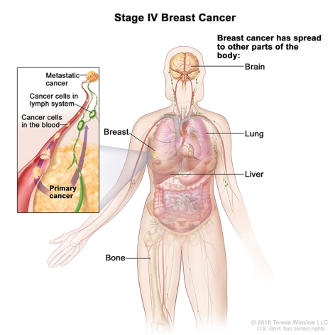
Stage IV (Metastatic)
The tumor can be any size and has spread to other organs, such as the bones, lungs, brain, liver, distant lymph nodes, or chest wall (any T, any N, M1). Metastatic breast cancer that is found when the cancer is first diagnosed is called de novo metastatic breast cancer. Most commonly, metastatic breast cancer is found after a previous diagnosis and treatment of early stage breast cancer.
Recurrent Breast Cancer
Recurrent cancer is cancer that has come back after treatment. Even when the cancer seems to be completely destroyed, the disease sometimes returns because undetected cancer cells remain somewhere in your body after treatment. It may return to the breast or chest wall. Or it may return in any other part of the body, such as the bones, liver, lungs, or brain. If the cancer does return, there will be another round of tests — similar to those done at the time of the original diagnosis — to learn about the extent of the recurrence.
Breast Cancer Tumor Grade
Cancer cells are given a grade when they are removed from the breast and checked in the lab. The grade is based on how much the cancer cells look like normal cells and gives a measurement of how aggressive the cancer cells appear to be. The information gives the oncologist a better idea of how quickly or slowly the cancer will grow and spread.
The invasiveness of breast cancer is evaluated and then given a score. Cancer that looks similar to healthy tissue and has different cell groupings, it is called "well-differentiated" or a "low-grade tumor." Cancerous tissue that looks very different from healthy tissue is called "poorly differentiated" or a "high-grade tumor."
-
Grade 1 or well differentiated (Nottingham score 3, 4, or 5). The cells are slower-growing, and look more like normal breast cells.
-
Grade 2 or moderately differentiated Nottingham (score 6, 7). The cells are growing at a speed of and look like cells somewhere between grades 1 and 3.
-
Grade 3 or poorly differentiated (Nottingham score 8, 9). The cancer cells look very different from normal cells and will probably grow and spread faster.